Brain Imaging in Diagnosing Autism is an evolving field that promises to enhance early detection, improve our understanding of the neurological roots of autism, and personalize treatment plans.
With the increasing global prevalence of autism spectrum disorder (ASD), researchers and clinicians are turning to advanced technologies like MRI, fMRI, and EEG to decode the brain’s complex architecture and function.
This article explores how these brain imaging tools contribute to diagnosing autism and what the future may hold.
🔍 What is Brain Imaging?
Brain imaging refers to a variety of technologies that capture images or signals from the brain to study its structure or activity. It can be broadly categorized into two types:
- Structural Imaging – focuses on brain anatomy (e.g., MRI)
- Functional Imaging – captures real-time brain activity (e.g., fMRI, EEG)
These tools are increasingly used in autism research to identify subtle but consistent brain differences between neurotypical and autistic individuals.
🧬 Why Brain Imaging Matters in Autism Diagnosis
Traditionally, autism is diagnosed through behavioral assessments, such as the ADOS (Autism Diagnostic Observation Schedule) and interviews. While effective, these methods are subjective and sometimes result in delayed diagnoses. Brain imaging offers:
- Objective biomarkers
- Early detection before behavior manifests
- Deeper insights into individual brain development
This technological approach could revolutionize personalized autism treatment and shorten the diagnostic timeline.
🧲 Magnetic Resonance Imaging (MRI)
MRI provides detailed images of brain structures. Studies using MRI have shown:
- Differences in brain volume in regions like the amygdala, which regulates emotion
- Altered growth trajectories in the brains of young autistic children
- Variations in white matter connectivity, especially in the corpus callosum
These structural differences may not be visible to the naked eye but can be crucial markers when interpreted by specialists.
📖 Study Spotlight: A 2017 study from UNC Chapel Hill used MRI scans at 6 and 12 months of age to predict autism diagnosis by age 2 with up to 80% accuracy. (NIH source)
🔄 Functional MRI (fMRI)
fMRI tracks brain activity by measuring changes in blood flow. It’s been particularly useful in identifying:
- Social brain network abnormalities
- Reduced synchronization in brain regions responsible for social communication
- Altered response to faces and social cues
This can help researchers understand the neurobiological basis of social difficulties in autism.
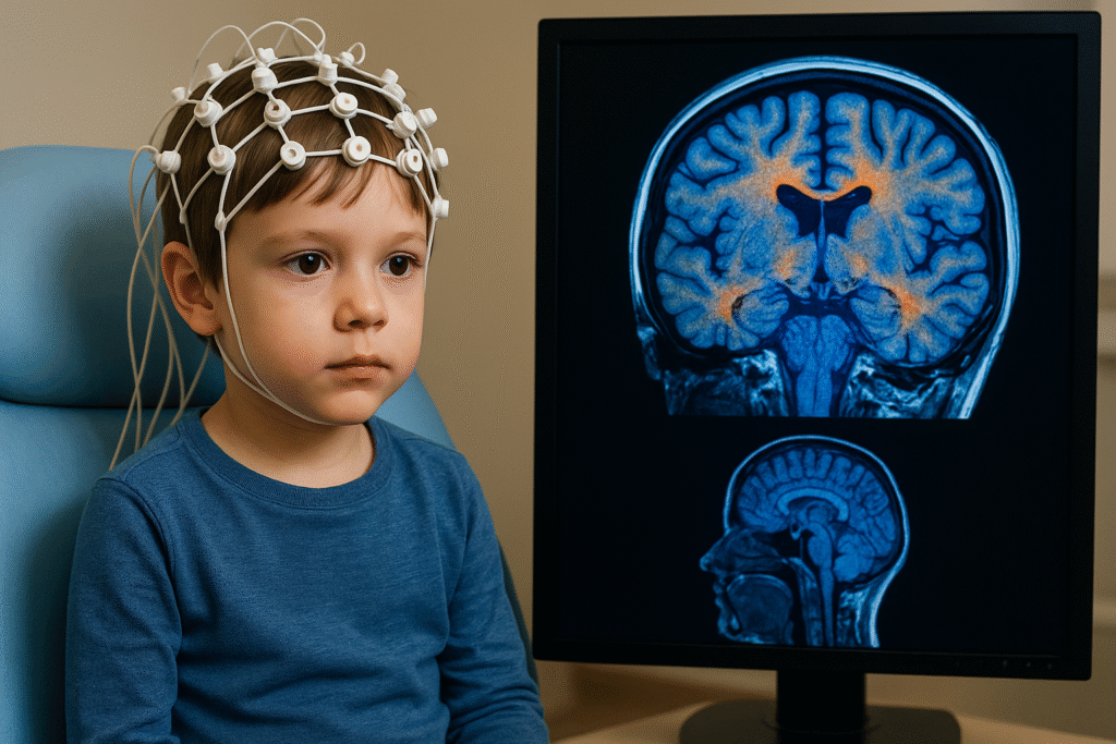
⚡ Electroencephalography (EEG)
EEG is a non-invasive method that records electrical activity via scalp electrodes. Its advantages include being:
- Affordable
- Quick
- Child-friendly
EEG studies have discovered:
- Slower brainwave patterns in response to stimuli
- Impaired sensory processing signals
- Reduced neural connectivity
Some researchers propose that EEG could be used as a clinical screening tool in pediatric populations.
🧪 PET and MEG: Emerging Tools
While less commonly used due to their complexity, PET (Positron Emission Tomography) and MEG (Magnetoencephalography) provide valuable insight:
- PET shows metabolic activity, revealing how brain cells process energy
- MEG captures magnetic fields produced by neuronal activity, offering high-resolution timing data
These methods are still primarily used in research settings, but their findings may guide future diagnostic protocols.
📊 Comparative Table of Imaging Methods
| Imaging Type | Focus | Pros | Cons |
|---|---|---|---|
| MRI | Brain structure | High resolution, non-invasive | Expensive, requires stillness |
| fMRI | Brain activity | Functional data, spatial accuracy | Time-consuming, less child-friendly |
| EEG | Brain activity | Portable, low cost, child-compatible | Low spatial resolution |
| PET | Metabolic activity | Unique insights into brain function | Uses radiation, costly |
| MEG | Neural timing | Excellent temporal resolution | Requires special equipment |
🧠 What Has Brain Imaging Revealed About Autism?
Some consistent findings across studies include:
- Hyperconnectivity or hypoconnectivity in certain brain regions
- Altered mirror neuron system, potentially explaining empathy challenges
- Disrupted default mode network, linked to self-referential thinking
These discoveries suggest that autism is not caused by a single “autism gene” or one brain defect. Instead, it reflects complex, whole-brain network changes.
🧩 Can Brain Imaging Replace Behavioral Diagnosis?
Not yet. Despite its promise, brain imaging is not ready to replace traditional assessments because:
- Findings are not uniform across all individuals
- Scanning methods are costly and time-consuming
- Results often need expert interpretation
However, combining imaging with behavioral data can enhance diagnostic accuracy, especially in complex or early-stage cases.
🧬 The Future of Autism Diagnosis with Brain Imaging
Technological advancements like AI-based image analysis and machine learning are now being used to detect patterns invisible to human eyes. These innovations may lead to:
- Automated early diagnosis tools
- Neurodiversity-informed brain mapping
- Tailored therapy recommendations
🔗 Internal & External Resources
- Autism and Sensory Processing Disorder: What’s the Difference?
- Early Signs of Autism in Toddlers Every Parent Should Know
- NIH Brain Imaging in Autism Research (DoFollow)
- Autism Speaks: Research on Brain Development (DoFollow)
- Brain Imaging Techniques – NIMH (DoFollow)
🧩 Final Thoughts on Brain Imaging in Diagnosing Autism
Brain Imaging in Diagnosing Autism is not science fiction—it’s already reshaping how we think about early detection and personalized interventions.
While it’s not yet a standalone diagnostic tool, brain imaging enhances our understanding of autism’s roots and could soon make diagnoses faster, more accurate, and more inclusive.
As research continues, the fusion of neurotechnology and behavioral science may finally unlock the secrets of the autistic brain.